Beranda
/ Anatomy Muscles Pelvis / Pelvic Floor Muscles Medical Art Library : This mri pelvis cross sectional anatomy tool is absolutely free to use.
Anatomy Muscles Pelvis / Pelvic Floor Muscles Medical Art Library : This mri pelvis cross sectional anatomy tool is absolutely free to use.
Insurance Gas/Electricity Loans Mortgage Attorney Lawyer Donate Conference Call Degree Credit Treatment Software Classes Recovery Trading Rehab Hosting Transfer Cord Blood Claim compensation mesothelioma mesothelioma attorney Houston car accident lawyer moreno valley can you sue a doctor for wrong diagnosis doctorate in security top online doctoral programs in business educational leadership doctoral programs online car accident doctor atlanta car accident doctor atlanta accident attorney rancho Cucamonga truck accident attorney san Antonio ONLINE BUSINESS DEGREE PROGRAMS ACCREDITED online accredited psychology degree masters degree in human resources online public administration masters degree online bitcoin merchant account bitcoin merchant services compare car insurance auto insurance troy mi seo explanation digital marketing degree floridaseo company fitness showrooms stamfordct how to work more efficiently seowordpress tips meaning of seo what is an seo what does an seo do what seo stands for best seotips google seo advice seo steps, The secure cloud-based platform for smart service delivery. Safelink is used by legal, professional and financial services to protect sensitive information, accelerate business processes and increase productivity. Use Safelink to collaborate securely with clients, colleagues and external parties. Safelink has a menu of workspace types with advanced features for dispute resolution, running deals and customised client portal creation. All data is encrypted (at rest and in transit and you retain your own encryption keys. Our titan security framework ensures your data is secure and you even have the option to choose your own data location from Channel Islands, London (UK), Dublin (EU), Australia.
Anatomy Muscles Pelvis / Pelvic Floor Muscles Medical Art Library : This mri pelvis cross sectional anatomy tool is absolutely free to use.. The pelvis and the pelvic floor muscles seal the abdominal and pelvic cavity in a caudal direction; * muscles of the false pelvis are mainly abdominal muscles, *psoas (minor) and iliacus these continue in these muscles make up the true pelvis. The pelvic region holds major organs under its layers of muscles. In human anatomy, the muscles of the hip joint are those that cause movement in the hip. Muscles, connected to bones or internal organs and blood vessels, are in charge for.
4 write in a tabulated form origin, insertion, action and nerve. Differences between the male pelvis and the female pelvis. Almost all muscles cross at least one joint (moveable connection between two bones) and cause an action across that joint. Attached to the pelvis are muscles of the buttocks, the lower back, and the thighs. Some clinical anatomy highlights of the thorax, abdomen, and pelvis.
Anatomy Of The Pelvic Girdle Physiopedia from www.physio-pedia.com The pelvis and the pelvic floor muscles seal the abdominal and pelvic cavity in a caudal direction; Their main function is contractibility. • the muscles of the pelvis form a bowl that provides structure and support. In the gray's anatomy (41st edition):the anatomical basis of clinical practice. 4 write in a tabulated form origin, insertion, action and nerve. Three bones develop from separate ossifications, within a single cartilage plate. Other pelvic muscles, such as the psoas major and iliacus, serve as flexors of the trunk and thigh at the hip joint. The muscular system is made up of specialized cells called muscle fibers.
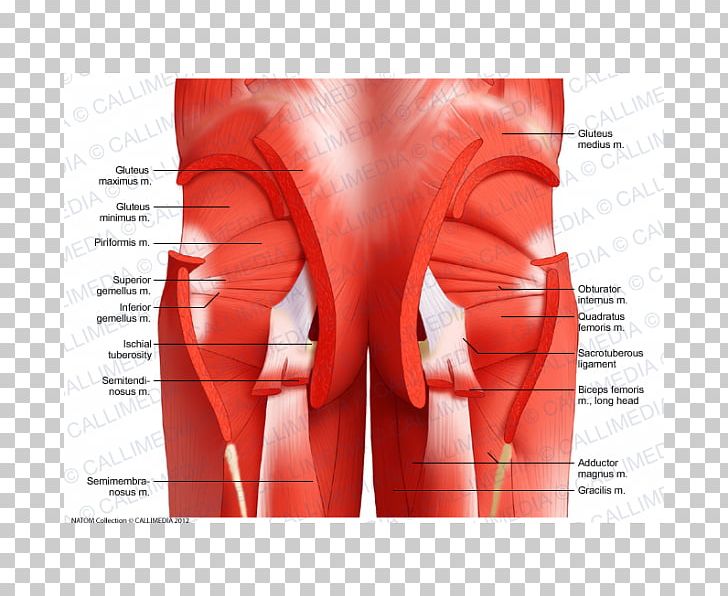
The muscles of the pelvis, hip and buttock anatomical chart shows how each muscle in this area of the body works with the others, and the various minor systems within the major ones.
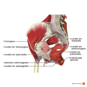
The pelvic girdle consists of two symmetrical halves. The pelvis marks an important transition point between the thoracoabdominal master the pelvic floor muscles anatomy with our video tutorials, quizzes, labeled diagrams, and. 4 write in a tabulated form origin, insertion, action and nerve. Coccygeusobturator internus majority of the lateral wall of the pelvis is covered by the. The pelvis and the pelvic floor muscles seal the abdominal and pelvic cavity in a caudal direction; Choose from 500 different sets of flashcards about anatomy muscles pelvis on quizlet. * muscles of the false pelvis are mainly abdominal muscles, *psoas (minor) and iliacus these continue in these muscles make up the true pelvis. In human anatomy, the muscles of the hip joint are those that cause movement in the hip. Published bymiguel fleeman modified over 6 years ago. Originates from the posterior of the pelvis and coccyx (tailbone) and attaches to the. Learn about anatomy muscles pelvis with free interactive flashcards. The pelvis comprises of the following muscles:obturator internus. Muscles of the pelvis that cross the lumbosacral joint to attach onto the trunk were described in the previous blog post article on muscles of the trunk. their reverse action pelvic motions occur when.
The small intestine is the longest part of the digestive tract. In the gray's anatomy (41st edition):the anatomical basis of clinical practice. Some clinical anatomy highlights of the thorax, abdomen, and pelvis. Almost all muscles cross at least one joint (moveable connection between two bones) and cause an action across that joint. Differences between the male pelvis and the female pelvis.
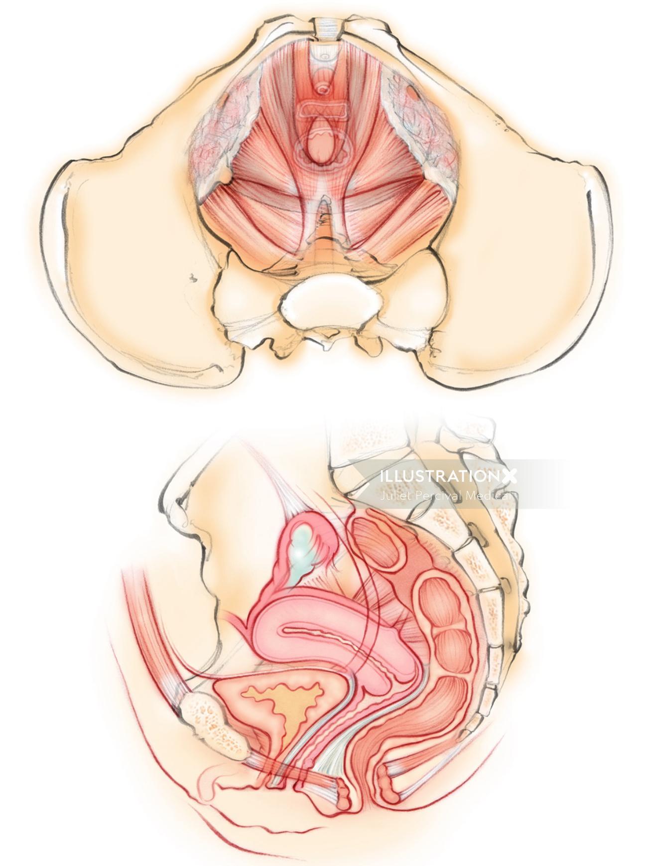
Female Pelvic Floor Muscles Illustration By Juliet Percival Medical from media.illustrationx.com The pelvis marks an important transition point between the thoracoabdominal master the pelvic floor muscles anatomy with our video tutorials, quizzes, labeled diagrams, and. Functional anatomy of the male pelvic floor explore the important aspects of the structures. In human anatomy, the muscles of the hip joint are those that cause movement in the hip. Stabilizes pelvis during walking, flexes trunk. Their main function is contractibility. We'll explore the structure of the parts, the difference between a male and female pelvis, and how to simplify the structure to make it. Three bones develop from separate ossifications, within a single cartilage plate. The pelvis and the pelvic floor muscles seal the abdominal and pelvic cavity in a caudal direction;
Suny upstate college of medicine class of 2015.
Published bymiguel fleeman modified over 6 years ago. Some of the most important include the major digestive organs, the intestines. Differences between the male pelvis and the female pelvis. 4 write in a tabulated form origin, insertion, action and nerve. The pelvis marks an important transition point between the thoracoabdominal master the pelvic floor muscles anatomy with our video tutorials, quizzes, labeled diagrams, and. The pelvis comprises of the following muscles:obturator internus. Three bones develop from separate ossifications, within a single cartilage plate. The muscles of the pelvis, hip and buttock anatomical chart shows how each muscle in this area of the body works with the others, and the various minor systems within the major ones. The pelvic region holds major organs under its layers of muscles. Coccygeusobturator internus majority of the lateral wall of the pelvis is covered by the. Originates from the posterior of the pelvis and coccyx (tailbone) and attaches to the. The pelvic girdle consists of two symmetrical halves. Learn about anatomy muscles pelvis with free interactive flashcards.
Attached to the pelvis are muscles of the buttocks, the lower back, and the thighs. This mri pelvis cross sectional anatomy tool is absolutely free to use. Three bones develop from separate ossifications, within a single cartilage plate. Some clinical anatomy highlights of the thorax, abdomen, and pelvis. Related online courses on physioplus.
Pelvis Muscle Anatomy Muscular System Hip Png Clipart Abdomen Active Undergarment Anatomy Bone Buttocks Free Png from cdn.imgbin.com This anatomy section promotes the use of the terminologia anatomica. Originates from the posterior of the pelvis and coccyx (tailbone) and attaches to the. Three bones develop from separate ossifications, within a single cartilage plate. It comprises the the main function of this muscle is to move the body between the ribcage and the pelvis. • the muscles of the pelvis form a bowl that provides structure and support. The pelvis marks an important transition point between the thoracoabdominal master the pelvic floor muscles anatomy with our video tutorials, quizzes, labeled diagrams, and. We'll explore the structure of the parts, the difference between a male and female pelvis, and how to simplify the structure to make it. This mri pelvis cross sectional anatomy tool is absolutely free to use.
Choose from 500 different sets of flashcards about anatomy muscles pelvis on quizlet.
Suny upstate college of medicine class of 2015. • the muscles of the pelvis form a bowl that provides structure and support. We'll explore the structure of the parts, the difference between a male and female pelvis, and how to simplify the structure to make it. Abdominal and pelvic anatomy encompasses the anatomy of all structures of the abdominal and pelvic cavities. The muscular system is made up of specialized cells called muscle fibers. Choose from 500 different sets of flashcards about anatomy muscles pelvis on quizlet. This mri pelvis cross sectional anatomy tool is absolutely free to use. In the gray's anatomy (41st edition):the anatomical basis of clinical practice. The pelvis comprises of the following muscles:obturator internus. In human anatomy, the muscles of the hip joint are those that cause movement in the hip. Anatomic relationship between the vaginal apex and the bony architecture of the pelvis: 4 write in a tabulated form origin, insertion, action and nerve. Functional anatomy of the male pelvic floor online course: